Pelvic Anatomy Ligaments : Male Hip Bones And Ligaments Labeled Rear View On White Stock Photo Download Image Now Istock - The pelvic girdle and pelvic spine.
Pelvic Anatomy Ligaments : Male Hip Bones And Ligaments Labeled Rear View On White Stock Photo Download Image Now Istock - The pelvic girdle and pelvic spine.. The cardinal ligament is a paired thickening of the parametrium and pelvic fascia at the base of the broad ligament, which extends between the cervix and vaginal fornix medially to the sidewall of pelvis laterally. Shop our carefully curated collection of high quality products today! The enclosed space between the inlet and outlet is called the true pelvis, with. They form what can be described as a basket weave formation, in order to create strength and tensegrity within the structure. The sacrospinous and sacrotuberous ligaments contribute to the formation of the greater and lesser sciatic foramens.
The sacrospinous and sacrotuberous ligaments contribute to the formation of the greater and lesser sciatic foramens. Other ligaments attached to bony pelvis include the sacrococcygeal ligaments, pubic symphysis ligaments, and endopelvic fascia ligament. Two innominate bones, which consist of the: The pelvic girdle and pelvic spine. The broad ligament is subdivided into the following:
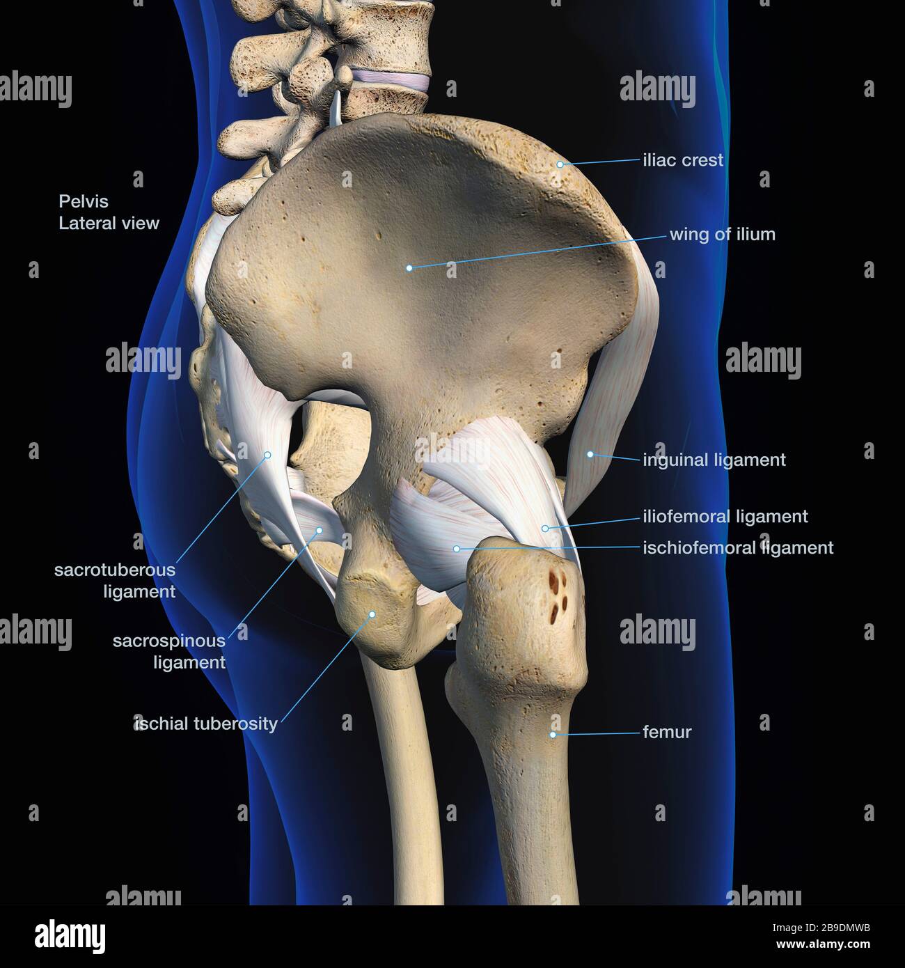
Lateral View Of Male Pelvis Hip Leg Bones And Ligaments On Black Background Stock Photo Alamy from c8.alamy.com This is part of the forced closure method that the pelvis adopts in order to keep itself secure. The suspensory ligament of the ovary, also infundibulopelvic ligament (commonly abbreviated ip ligament or simply ip), is a fold of peritoneum that extends out from the ovary to the wall of the pelvis. The sacrospinous ligament spans the sacrum to the ischial spine, and the sacrotuberous ligament spans the sacrum to the ischial tuberosity. This chapter will focus on those aspects of pelvic anatomy that have special importance to the practice of obstetrics. It is usually divided into two separate anatomic regions: The pelvic girdle, also known as the hip bone, is composed of three fused bones: Ligaments of the pelvis and hip. Cardinal ligament and the uterosacral ligaments provide apical support for the uterus and upper vagina.
All axis rotatable modeled 3d model pelvis and ligaments, labeled.
This image shows the posterior back view of the female pelvic brim (the bones and ligaments that forms the pelvic region in the female) showing: The broad ligament folds over the fallopian tubes and ovaries and covers them anteriorly and posteriorly. • anterolateral wall—hip bone and obturator internus muscles. These ligaments are important stabilizers. We hope you can get the exact. Imaios and selected third parties, use cookies or similar technologies, in particular for audience measurement. • also known as pelvic cavity. • lateral boundaries—fused ilium and ischium. The named ligaments of the pelvis mostly arise from the sacrum and attach to varying segments of the pelvic bone. The ligaments, made of strong connective tissue, which connect bones to bones, and the tendons, which connect muscles to bones. The outlet is formed by the pubic arch, ischial spines, sacrotuberous ligaments, and the coccyx. The sacral ligaments are responsible for the major connection between the three bones of the pelvis. Bones and ligaments of the female pelvis.
In addition to the labrum and the ligamentum teres, three other. The sacral ligaments are responsible for the major connection between the three bones of the pelvis. The femoral ligaments act to stabilize the ball and socket joint of the hip, connecting to the ilium and the ischium. Broad ligament the broad ligament supports the uterus, fallopian tubes, and ovaries. Ligaments attach the lateral border of the sacrum to various bony landmarks on the bony pelvis to aid stability.
Male Pelvic Model 7 Part from cdn11.bigcommerce.com The ligaments of the pelvis, are amongst the strongest in the human body. The pelvic ligaments are strong, thick bands of fibrous tissue that connect the pelvic bones. Lets get deeper into the musculoskeletal anatomy of the hip and look at the bones and bony bits of the pelvis, and the ligaments that attach here and hold it. Broad ligament the broad ligament supports the uterus, fallopian tubes, and ovaries. The ligaments, made of strong connective tissue, which connect bones to bones, and the tendons, which connect muscles to bones. The broad ligament is a flat sheet of peritoneum, associated with the uterus, fallopian tubes and ovaries. This is part of the forced closure method that the pelvis adopts in order to keep itself secure. The pelvis is held together by three principal ligaments:
Thank you for visit anatomynote.com.
• muscles and ligaments form a pelvic floor. The pelvis is held together by three principal ligaments: The femoral ligaments act to stabilize the ball and socket joint of the hip, connecting to the ilium and the ischium. The sacrospinous ligament spans the sacrum to the ischial spine, and the sacrotuberous ligament spans the sacrum to the ischial tuberosity. The ligaments of the pelvis, are amongst the strongest in the human body. • posterolateral wall—piriformis and coccygeus muscles. This image added by admin. Broad ligament the broad ligament supports the uterus, fallopian tubes, and ovaries. Ligaments connect one bone to another and provide important stability. The inlet to the pelvic canal is at the level of the sacral promontory and superior aspect of the pubic bones.; Imaios and selected third parties, use cookies or similar technologies, in particular for audience measurement. Shop our carefully curated collection of high quality products today! Some sources consider it a part of the broad ligament of uterus while other sources just consider it a termination of the ligament.
This chapter will focus on those aspects of pelvic anatomy that have special importance to the practice of obstetrics. The pelvis is a boney structure at the base of the lumbar spine. It extends from the lateral pelvic walls on both sides, and folds over the internal female genitalia, covering their surface anteriorly and posteriorly. Iliolumbar, sacrotuberous and sacrospinous ligaments. Joints and ligaments of the pelvis (anterior view)
Amazon Com Somso Ms8 1 Female Pelvis Model With Vessels Ligaments Muscles Nerves Female Organs Industrial Scientific from m.media-amazon.com The outlet is formed by the pubic arch, ischial spines, sacrotuberous ligaments, and the coccyx. Joints and ligaments of the pelvis the two sacroiliac joints are synovial joints, and are further strengthened by the very strong posterior sacroiliac ligaments which run along the posterior aspect of the joint. • posterolateral wall—piriformis and coccygeus muscles. Ligaments of the pelvis the posterior sacroiliac ligament supports the sacroiliac joint. The pelvis is a boney structure at the base of the lumbar spine. Some sources consider it a part of the broad ligament of uterus while other sources just consider it a termination of the ligament. Ligaments connect one bone to another and provide important stability. Broad ligament the broad ligament supports the uterus, fallopian tubes, and ovaries.
The broad ligament folds over the fallopian tubes and ovaries and covers them anteriorly and posteriorly.
Also, the compartmental anatomy of the female pelvis is explained, including the extraperitoneal pelvic spaces. The sacrospinous ligament spans the sacrum to the ischial spine, and the sacrotuberous ligament spans the sacrum to the ischial tuberosity. The broad ligament is subdivided into the following: The named ligaments of the pelvis mostly arise from the sacrum and attach to varying segments of the pelvic bone. The pelvis itself is a paired composite structure made up by three bones (ilium, ischium and pubis) that articulates with the sacral part of the axial spine. The broad ligament folds over the fallopian tubes and ovaries and covers them anteriorly and posteriorly. This is part of the forced closure method that the pelvis adopts in order to keep itself secure. Cardinal ligament and the uterosacral ligaments provide apical support for the uterus and upper vagina. The inlet to the pelvic canal is at the level of the sacral promontory and superior aspect of the pubic bones.; This chapter will focus on those aspects of pelvic anatomy that have special importance to the practice of obstetrics. Thank you for visit anatomynote.com. The suspensory ligament of the ovary, also infundibulopelvic ligament (commonly abbreviated ip ligament or simply ip), is a fold of peritoneum that extends out from the ovary to the wall of the pelvis. The enclosed space between the inlet and outlet is called the true pelvis, with the plane of the inlet being at right angles to the plane of the outlet.
Ligaments connect one bone to another and provide important stability pelvic anatomy. Finally, a checklist is provided for structured reporting of the mri findings in the female pelvis.


0 Komentar